Why teaching AI about symmetry could make prenatal diagnosis more reliable

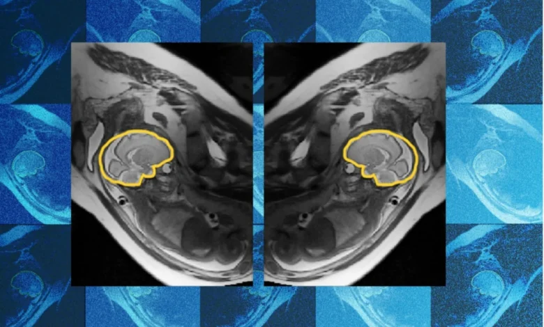
Despite millions of years of human evolutionary refinement, the gestational development of the human fetus remains an extraordinarily intricate and vulnerable biological process. Susceptible to a myriad of genetic, environmental, and metabolic disruptions, prenatal life can occasionally veer off course, giving rise to developmental abnormalities. Among the earliest bodily systems to form—and one that continues its complex maturation long after birth—is the human brain. Identifying structural or functional deviations in utero has long stood as a paramount challenge in modern obstetrics and pediatric neurology, largely due to the physical limitations of prenatal imaging technologies.
Traditionally, when routine prenatal ultrasounds reveal potential anomalies in a developing fetal brain, sonographers flag the cases for follow-up magnetic resonance imaging (MRI). While MRI provides superior soft-tissue contrast compared to ultrasound, the technology faces a formidable barrier: relentless, unpredictable fetal movement. Because a fetus constantly shifts, tumbles, and floats inside the amniotic fluid, capturing a clear, diagnostically reliable image has historically relied on manual adjustments, patient endurance, and a significant margin of guesswork. Imperfect imaging frequently leads to missed diagnoses or, conversely, false alarms that heighten parental anxiety and complicate clinical decision-making.
To bridge this technological gap, an interdisciplinary team of researchers hailing from the Massachusetts Institute of Technology (MIT), Boston Children’s Hospital, and the French research institute Inria has engineered a pioneering deep learning model known as E(3)-Pose. Designed to track and image a fetus’s head dynamically in real-time while it is in motion, the model reconstructs high-resolution composite images that radiologists can rigorously examine for pathologies. The breakthrough research behind E(3)-Pose was formally presented at the 2026 European Conference on Computer Vision, marking a significant milestone in medical imaging artificial intelligence.
The Physics of Motion: Overcoming the 2D "Slice" Problem
To understand the magnitude of the engineering hurdle E(3)-Pose clears, one must examine how a standard fetal MRI functions. Unlike a photograph captured in a fraction of a second, an MRI scanner acquires data sequentially, generating a series of individual two-dimensional (2D) planes, or "slices." When stacked together through computational processing, these 2D slices are intended to form a cohesive, high-resolution three-dimensional (3D) volume of the fetal head.
However, fetal movement shatters this geometric assumption. "One of the problems with having the baby move is that the slices are all over the place," explains Ramya Muthukrishnan, the lead author of the study and an MIT doctoral student working within the lab of Polina Golland. Golland serves as the Sunlin (1966) and Priscilla Chou Professor of Electrical Engineering and Computer Science at MIT, alongside roles as a principal investigator at the MIT Abdul Latif Jameel Clinic for Machine Learning in Health (Jameel Clinic) and the MIT Computer Science and Artificial Intelligence Laboratory (CSAIL).
When a fetus shifts mid-scan, consecutive 2D slices are inevitably captured from disparate, misaligned angles. This spatial dislocation creates coverage gaps—blind spots in the volumetric data—and unusual slice orientations. "There are very fine details that you need a very good volumetric picture of the brain to see," Muthukrishnan notes. "If you have coverage gaps, you can’t see critical regions, and you need to be able to see well to detect abnormal development."
In clinical settings, these artifacts can obscure subtle microstructural anomalies, ventricular enlargement, cortical malformations, or early signs of hypoxic-ischemic injury. The resulting diagnostic uncertainty often forces physicians to request repeat scans, which extends the duration of clinical visits, strains hospital resources, and places physical discomfort on pregnant patients who must remain motionless inside a loud, confining machine for extended periods.
Baking Physics into Neural Network Architecture
What distinguishes E(3)-Pose from conventional computer vision models is its foundational reliance on physical principles rather than purely data-driven pattern recognition. Traditional machine learning models are typically fed vast datasets and left to infer physical laws empirically. In contrast, E(3)-Pose integrates the fundamental laws of physics directly into its neural network architecture.
Specifically, the model embeds the intrinsic left-right symmetry of the fetal head straight into its structural design. By hardcoding these geometric and physical constraints, E(3)-Pose dramatically reduces the model’s margin of error, enabling it to accurately identify, track, and predict the brain’s orientation even when movement is erratic and data inputs are noisy.
The path to developing this sophisticated system was far from straightforward. When Muthukrishnan embarked on her doctoral journey, her academic background was rooted firmly in computer science, with virtually no prior experience in physics or MRI hardware mechanics. Translating theoretical neural network designs into functional code capable of interfacing directly with high-powered hospital scanners proved to be a formidable baptism by fire.
"I was surprised when I had to go and implement this on the scanner," she recalls. "I was not prepared for how many times I would crash the scanner, because putting things on hardware and getting it to work on hardware is a different beast than training a model to run on data on a GPU."
A Collaborative Clinical Genesis
Muthukrishnan’s resolve was forged early in her PhD program while she was shadowing a pediatric radiologist performing fetal MRIs at Boston Children’s Hospital. Witnessing firsthand the shared frustration of patients and clinicians proved to be a transformative experience. Expectant mothers, often exhausted and uncomfortable after enduring an hour inside an MRI suite, would frequently reach their physical limit and have to abandon the scan halfway through. Simultaneously, radiologists were left staring at incomplete, motion-degraded datasets, unable to gather the definitive diagnostic evidence required to monitor fetal neurological maturation.
"Seeing frustration about it helped me decide what I wanted to do with my PhD, because it felt like this was a pain point," Muthukrishnan states.
Fortunately, she did not tackle the challenge in isolation. She was supported by a multidisciplinary roster of clinical and engineering experts. Co-author Borjan Gagoski, an assistant professor of radiology at Harvard Medical School and a researcher at Boston Children’s Hospital, engineered a specialized MRI acquisition sequence designed specifically to gather the secondary data required to train E(3)-Pose. Gagoski also worked closely with Muthukrishnan to troubleshoot and reboot hospital hardware whenever her experimental code caused scanner crashes.
Meanwhile, Aryn Lee, a research assistant at Boston Children’s Hospital, coordinated the recruitment of pregnant volunteers and facilitated the acquisition of the MRI scans essential for the project’s empirical validation. The research team was further bolstered by senior guiding figures, including Elfar Adalsteinsson, an MIT professor of electrical engineering and computer science; Ellen Grant, the Käthe Beutler Professor of Pediatrics and professor of radiology at Harvard Medical School; and Polina Golland. Benjamin Billot, a researcher at Inria and a former postdoctoral fellow in Golland’s laboratory, provided critical initial guidance, helping Muthukrishnan build upon foundational methodologies he had previously developed.
Clinical Endorsement and Expert Perspectives
The convergence of advanced mathematical modeling, rigorous hardware integration, and deep clinical insight has drawn widespread praise from medical imaging leaders.
"Ramya’s work elegantly combines geometric concepts like equivariance and symmetry with the power of deep learning to change how MRI scanners acquire the images, and to ultimately help the physicians detect and treat problems early in development," remarks Polina Golland. "It’s an impressive research project that is deep and fundamental in how it models image acquisition, with clear clinical impact."
Dr. Ellen Grant, who directs the Fetal Neonatal Neuroimaging and Developmental Science Center (FNNDSC) at Boston Children’s Hospital, emphasizes the transformative operational benefits E(3)-Pose promises for clinical workflows. According to Grant, Muthukrishnan’s innovations "will automate fetal brain MRI, replacing today’s slow, manual process with faster and more comfortable scans for mothers, while also providing higher-quality images for radiologists to interpret."
By reducing the need for lengthy, repeated scan sequences, the technology directly addresses patient compliance issues. Shorter, more efficient imaging sessions alleviate physical strain on expectant mothers while slashing the operational bottlenecks that frequently backlog pediatric radiology departments.
Beyond the Womb: Broader Implications and Applications
While the immediate clinical utility of E(3)-Pose is centered on prenatal neuroimaging, the underlying technological framework possesses expansive potential across a variety of disparate fields. Because the model excels at parsing noisy, high-dimensional spatial data from moving subjects using limited sensor inputs, its core principles can be readily adapted to image other anatomically complex or dynamic regions of the human body, such as cardiac structures or joint mechanics during motion.
Intriguingly, the engineering solutions developed for E(3)-Pose share strong conceptual parallels with challenges found in autonomous vehicle technology. Self-driving cars rely on limited arrays of sensors—such as cameras and LiDAR—to capture noisy, real-time 3D data in order to track moving objects in dynamic environments. The geometric equivariance and physics-informed architecture pioneered by the MIT-Boston Children’s team could theoretically enhance machine vision systems in robotics and automated transport, demonstrating how medical research frequently yields cross-industry technological dividends.
Future Horizons: Targeting Second-Trimester Vulnerabilities
Despite its success with third-trimester imaging data, the research team is not resting on its laurels. Muthukrishnan and her colleagues are actively working to refine and expand E(3)-Pose’s capabilities so that it can effectively image second-trimester fetuses or even younger gestational stages.
Adapting the model for earlier developmental stages presents a unique set of biological and technical hurdles. Second-trimester fetuses are significantly smaller than their third-trimester counterparts and possess considerably more room to move within the amniotic sac. Consequently, their motion is much more erratic, frequent, and difficult to predict.
Yet, overcoming this challenge remains an urgent priority for the medical community. "They’re more important to focus on because the earlier you detect brain abnormalities, the easier it is to intervene," Muthukrishnan stresses. Earlier detection opens critical windows for therapeutic interventions, parental counseling, and multidisciplinary birth planning, potentially altering the lifelong neurological trajectory of affected children.
Financial and institutional backing for the ongoing development of E(3)-Pose has been robustly provided by a coalition of premier research funds and government agencies, including the MIT Jameel Clinic, the MIT Takeda Program, the Chou Family Transformative Research Fund, the National Institute of Biomedical Imaging and Bioengineering, and the Eunice Kennedy Shriver National Institute of Child Health and Human Development.
As the research transitions further from academic prototyping toward eventual clinical translation and commercialization, E(3)-Pose stands as a testament to the power of interdisciplinary collaboration—bridging the gap between theoretical computer science, hospital hardware, and clinical empathy to illuminate the most delicate stages of human life.
Related posts: