Revolutionary AI System Developed by MIT Researchers Bridges 2D X-Rays and 3D Medical Scans to Transform Minimally Invasive Surgery

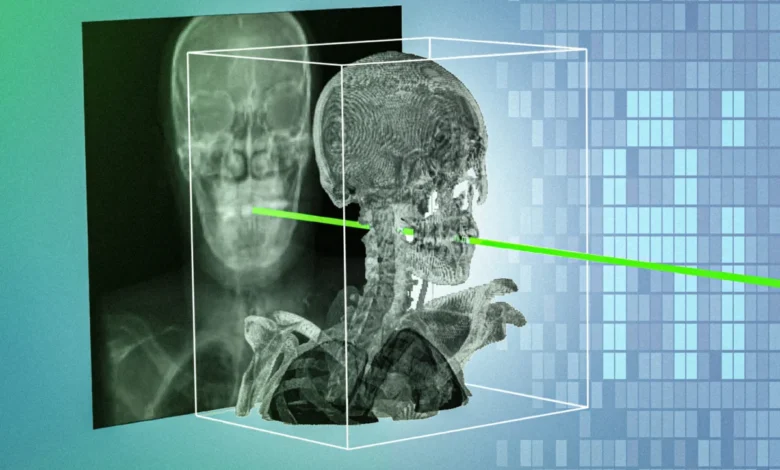
In modern medicine, minimally invasive procedures have long represented a triumph of technological refinement, allowing surgeons to treat complex conditions through tiny incisions with the aid of real-time imaging. Yet, a fundamental limitation has persisted in the operating room: the challenge of navigating inside the human body using flat, two-dimensional X-rays while trying to map those views against a patient’s rich, three-dimensional preoperative scans. This persistent disconnect increases procedure times, demands decades of specialized clinical training to master, and elevates the risk of procedural complications.
To resolve this critical bottleneck, a team of researchers and clinicians at the Massachusetts Institute of Technology (MIT) and collaborating medical institutions has engineered a breakthrough artificial intelligence system. Dubbed xvr—which stands for X-ray volume registration—the new technology rapidly and accurately matches intraoperative 2D X-rays with preoperative 3D medical images, such as computed tomography (CT) scans or magnetic resonance imaging (MRI) data. Operating with sub-millimeter precision in a matter of seconds, xvr represents a paradigm shift in surgical navigation, promising to make advanced, life-saving interventions faster, safer, and significantly more accessible to hospitals outside major metropolitan medical centers.
The details of this groundbreaking development were published today in the journal Nature, marking a major milestone in medical computer vision and clinical engineering.
The Persistent Challenge of 2D/3D Registration in Surgery
To understand the magnitude of the MIT team’s achievement, one must examine the daily realities of modern interventional radiology and surgery. During procedures such as angioplasty to clear obstructed arteries, cardiac catheterizations, or emergency interventions for acute ischemic strokes, clinicians rely heavily on fluoroscopy—real-time mobile X-ray imaging—to track the trajectory of micro-instruments like catheters, guidewires, and endoscopes.
However, because X-ray imaging projects a three-dimensional volume onto a flat, two-dimensional plane, depth perception is inherently lost. The resulting images are grainy and require immense spatial reasoning to interpret. To compensate for this limitation, physicians frequently attempt to align the live X-ray stream with the patient’s preoperative 3D scans—a complex geometric alignment process known in medical imaging as 2D/3D registration.
Historically, this alignment has been performed manually. A clinician must scrutinize the displays, guess the precise coordinates of a surgical tool, and manually input numerical parameters or click on anatomical landmarks on a monitor. This manual workflow is notoriously slow, burdensome, and prone to human error, consuming valuable minutes during high-stress procedures where every second counts.
While researchers have previously attempted to automate this process using conventional machine learning and deep learning tools, these efforts have largely stumbled. Human anatomical diversity is vast; a neural network trained to recognize vascular structures or bone geometries in one patient often fails catastrophically when presented with the unique anatomical variations of another. Furthermore, the scarcity of large, high-quality, annotated medical datasets has made it exceedingly difficult to train generalized deep-learning models robust enough to handle the infinite variability of the human body across diverse clinical scenarios.
A New Approach: Patient-Specific Machine Learning and Physics Simulations
Faced with the limitations of generalized AI models, Vivek Gopalakrishnan—a postdoctoral researcher in the MIT Computer Science and Artificial Intelligence Laboratory (CSAIL) and lead author of the study—alongside his colleagues, pivoted to an entirely different computational paradigm. Instead of trying to build a single, universal machine learning model capable of handling every human anatomy, the team designed a framework focused on hyper-customization for the individual patient.
"We tailor this one specific model for this one specific patient, and it doesn’t matter if it works on other people because there will be different models for those people," Gopalakrishnan explains.
The xvr framework initiates this customization process by taking a patient’s preoperative 3D scan—whether a high-resolution CT or MRI—and feeding it into a rigorous, physics-based simulator. Rather than using generative AI to fabricate synthetic pixels from scratch, which can introduce dangerous structural "hallucinations" or artifacts, the xvr simulator models the exact physical interaction of X-rays passing through human tissue.
In a matter of seconds, the system generates roughly 1,000 realistic, synthetic X-ray projections from a wide multitude of virtual angles. This massive repository of patient-specific, physics-grounded data is then utilized to rapidly train a customized AI registration model tailored exclusively to that individual’s unique internal geography.
Overcoming the Time Barrier with Foundation Models
While training a machine learning model from scratch for each individual patient yields exceptional, sub-millimeter accuracy, the computational overhead required is immense. Historically, training such a specialized neural network from the ground up would take approximately 12 hours—an operational impossibility in emergency surgical environments where medical decisions must be made instantaneously.
To solve this temporal paradox, the MIT team engineered a hybrid solution leveraging a high-capacity foundation model. The researchers gathered a vast, diverse repository of whole-body 3D medical scans encompassing more than 2,000 patients. This expansive dataset spanned a wide spectrum of ages—including both pediatric and adult cases—various imaging modalities, and numerous anatomical regions.
Using the xvr framework, the researchers leveraged these diverse scans to pretrain a versatile foundation model. When a new patient arrives at the hospital, this pretrained model does not need 12 hours of training; instead, it draws upon its foundational training to rapidly adapt to the new patient’s anatomical data in approximately five minutes.
Once adapted, the model can automatically register real-time intraoperative X-rays with the patient’s pre-existing 3D scans within seconds, achieving the exact same sub-millimeter precision as a model trained from scratch over half a day.
"So now you can get patient-specific accuracy but also in a very rapid time frame," Gopalakrishnan highlights.
Rigorous Validation Across Multiple Medical Centers
To prove the clinical viability of xvr, the research team subjected the framework to the largest and most rigorous evaluation dataset assembled for real 2D/3D medical registrations to date. The study incorporated multi-hospital data drawn from five distinct clinical institutions, encompassing dozens of unique bone structures, organ systems, and complex vascular networks across both adult and pediatric patient populations.
The results demonstrated that xvr significantly outperformed existing artificial intelligence registration methodologies by an entire order of magnitude. It maintained robust stability and high precision across a remarkably wide range of patient anatomies and medical specialties, proving its capability to operate efficiently under the stringent time constraints demanded by emergency interventions.
The breadth of expertise behind the study underscores its clinical relevance. In addition to lead author Vivek Gopalakrishnan, the research was co-led by senior authors Polina Golland, the Sunlin and Priscilla Chou Professor of Electrical Engineering and Computer Science at MIT, principal investigator in CSAIL, and leader of the Medical Vision Group; and Neel Dey, a former CSAIL postdoc who is now an investigator at Harvard Medical School and Massachusetts General Hospital.
The collaborative team also included clinical experts across several medical disciplines: David-Dimitris Chlorogiannis, a researcher and clinician at Harvard Medical School; Andrew Abumoussa, a neurosurgeon at St. Luke’s Marion Bloch Neuroscience Institute; Anna M. Larson, a pediatric clinician at Shriners Children’s Hospital; Nazim Haouchine, an assistant professor of radiology at Harvard and Brigham and Women’s Hospital; Darren B. Orbach, a physician and scientist at Boston Children’s Hospital; and Sarah Frisken, an associate professor of radiology at Harvard.
Broader Implications for Healthcare Access and Surgical Robotics
The successful translation of xvr from computational theory to validated medical tool carries profound implications for public health, healthcare equity, and the future of surgical robotics.
Emergency interventions—particularly for time-sensitive neurological events such as acute ischemic strokes—rely heavily on rapid access to specialized endovascular procedures. However, geographic disparities in healthcare infrastructure remain a persistent barrier.
"A majority of Americans live more than an hour away from a center that can perform noninvasive procedures, like emergency stroke interventions. An hour in stroke time is incredibly substantial," Gopalakrishnan points out. "Making these procedures easier by combining 2D and 3D information enables these types of highly specialized life-saving procedures to be more accessible to much broader parts of the population."
By reducing the cognitive load and technical complexity required to interpret flat 2D X-ray imagery, xvr could potentially democratize access to advanced minimally invasive interventions. Procedures that currently demand decades of specialized sub-board training could become standard protocols performable across a wider network of regional community hospitals, thereby shrinking the critical time-to-treatment window for rural and underserved patient populations.
Furthermore, the technology holds significant promise for the burgeoning field of robotic surgery. Modern surgical robots rely on precise spatial awareness to execute delicate maneuvers inside the human body. By providing real-time, sub-millimeter alignment between intraoperative tools and preoperative anatomical maps, xvr can serve as an advanced navigational backbone for autonomous or semi-autonomous surgical robotic platforms, enhancing both safety profiles and operational efficiency.
Looking Ahead: Commercialization and Real-Time Deployment
Following two years of meticulous algorithmic development, rigorous cross-institutional validation, and peer-reviewed publishing, the MIT-led research collective is now setting its sights on real-world clinical translation.
The immediate technical objectives for the research team include optimizing the xvr framework for even faster real-time deployment, conducting expanded clinical trials to verify operational reliability across an even wider array of emergency scenarios, and expanding the underlying algorithms to seamlessly account for dynamic, moving body parts such as beating hearts and expanding lungs.
At the same time, the team is actively forging commercial partnerships to transition the technology from an academic research project into tangible, FDA-cleared clinical software and hardware integrations.
"For the past two years, we’ve been carefully developing this algorithm and validating it," Gopalakrishnan concludes. "Now, we are collaborating closely with surgical robotics companies and clinical groups to turn this research into useful tools for navigation or deployment."
Financial support for the research was provided, in part, by the National Institutes of Health (NIH), the MIT CSAIL-Wistron Program, the MIT-IBM Computing Research Lab, the MIT Jameel Clinic, the MIT Health and Life Sciences Collaborative, and the Chou Family Transformative Research Fund.
Related posts: