Revolutionary AI Framework Bridges 2D X-Rays and 3D Scans to Transform Precision Surgery

In the high-stakes environment of minimally invasive surgery, a fraction of a millimeter can mean the difference between a successful intervention and a catastrophic complication. Surgeons guiding catheters, endoscopes, and specialized tools through microscopic incisions have long relied on real-time, two-dimensional X-rays to visualize their progress. However, translating a flat, grayscale image into a mental map of a patient’s complex, three-dimensional internal anatomy is an immense cognitive challenge that typically requires decades of rigorous medical training.
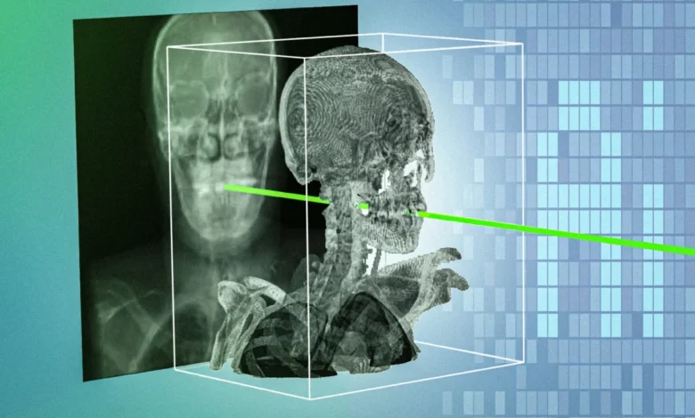
To solve this persistent limitation in modern medicine, an interdisciplinary team of researchers and clinicians at the Massachusetts Institute of Technology (MIT), alongside collaborating medical institutions, has developed a groundbreaking artificial intelligence framework known as xvr (X-ray volume registration). Published in the journal Nature, this novel technology accurately and rapidly aligns real-time surgical X-rays with a patient’s preoperative three-dimensional medical scans, such as CT scans and MRIs, achieving sub-millimeter precision in a matter of seconds. By bridging the gap between 2D intraoperative imaging and 3D preoperative planning, xvr promises to democratize access to complex, life-saving procedures, enhance surgical safety, and pave the way for more advanced robotic-assisted surgeries.
The Mechanics of Medical Image Registration
Minimally invasive procedures, ranging from routine angioplasties to open blocked arteries to emergency interventions for acute ischemic strokes, have transformed healthcare by reducing patient recovery times, lowering infection risks, and minimizing scarring. During these procedures, clinicians utilize mobile C-arm X-ray fluoroscopy units to generate live, high-speed images that allow them to monitor instruments inside the body.
Yet, because X-ray imaging projects a 3D volume onto a flat 2D plane, depth perception is inherently lost. Important anatomical structures can overlap, obscuring the precise location of a surgical tool relative to critical blood vessels, nerves, or delicate organs. To overcome this, clinicians historically practiced manual registration—a tedious and error-prone process where a physician must mentally estimate tool coordinates by entering numbers into a computer console or manually clicking anatomical landmarks on a monitor.
In recent years, computer scientists have attempted to streamline this workflow by developing deep-learning models capable of predicting 2D/3D registration automatically. However, these traditional machine-learning systems have repeatedly failed in clinical settings. Human anatomy is remarkably diverse; a generalized AI model trained on a standard dataset might perform exceptionally well for one patient while failing catastrophically for another. Furthermore, the scarcity of high-quality, densely annotated medical image datasets has severely limited the robustness of conventional deep-learning architectures, rendering them impractical for emergency departments and operating rooms.
A Paradigm Shift: Patient-Specific Machine Learning
Recognizing the limitations of universal AI models that attempt to fit every patient, the MIT research team pivoted toward a paradigm of patient-specific machine learning. Instead of designing a single model intended to work universally across all human bodies, the xvr framework constructs a customized model tailored explicitly to an individual patient using their own preoperative imaging data.
The workflow begins immediately after a patient undergoes a preoperative CT scan or MRI. The xvr system ingests this 3D scan and executes a high-speed, physics-based simulation of the X-ray imaging process. Rather than relying on generative AI techniques that synthesize data out of thin air—which can introduce dangerous hallucinations or fictional anatomical features—xvr strictly obeys the laws of physics. By simulating the passage of X-rays through the patient’s unique volumetric data from thousands of different angles, the system generates approximately 1,000 realistic synthetic X-ray images every second.
This massive repository of patient-specific synthetic data is then used to train an AI model to recognize and align the patient’s specific internal structures. While training such a model completely from scratch for every incoming patient would theoretically take up to 12 hours—an impossible timeframe for emergency interventions—the researchers engineered an innovative solution: a foundation model.
By leveraging whole-body 3D medical scans from more than 2,000 diverse patients encompassing various age groups, medical imaging modalities, and anatomical regions, the researchers pretrained a versatile base network using xvr. When applied to a new patient, this foundation model does not need to learn human anatomy from the ground up. Instead, it rapidly adapts to the individual’s specific scan in roughly five minutes, retaining the exact accuracy of a model trained over half a day. Once adapted, the model can automatically match intraoperative X-rays to the 3D scan in mere seconds.
Rigorous Validation Across Diverse Clinical Environments
To ensure the reliability and clinical viability of xvr, the research team subjected the framework to rigorous empirical testing using the largest available dataset of real-world 2D/3D registrations ever compiled. The evaluation incorporated retrospective data sourced from five distinct medical centers, spanning dozens of complex bone structures and organ systems across both adult and pediatric patient populations.
The results demonstrated that xvr significantly outperformed existing state-of-the-art AI registration methods by an order of magnitude. Its superior accuracy, coupled with its unprecedented robustness across varying patient anatomies and pathological conditions, established a new benchmark for medical image processing.
The research team behind this breakthrough represents a powerful collaboration between computational theorists and frontline medical practitioners. The project was led by Vivek Gopalakrishnan, a postdoc in the MIT Computer Science and Artificial Intelligence Laboratory (CSAIL) and a recent graduate of the Harvard-MIT Program in Health Sciences and Technology. Co-senior authors of the study include Polina Golland, the Sunlin and Priscilla Chou Professor of Electrical Engineering and Computer Science at MIT, principal investigator in CSAIL, and leader of the Medical Vision Group; and Neel Dey, an investigator at Harvard Medical School and Massachusetts General Hospital. Additional contributions were provided by a multidisciplinary roster of clinicians and researchers, including David-Dimitris Chlorogiannis of Harvard Medical School; neurosurgeon Andrew Abumoussa of the St. Luke’s Marion Bloch Neuroscience Institute; pediatric clinician Anna M. Larson of Shriners Children’s Hospital; assistant professor of radiology Nazim Haouchine of Harvard and Brigham and Women’s Hospital; physician-scientist Darren B. Orbach of Boston Children’s Hospital; and associate professor of radiology Sarah Frisken of Harvard.
Democratizing Access to Specialized Life-Saving Care
Beyond its technical achievements, the clinical implications of xvr extend deeply into public health and healthcare equity. Dr. Gopalakrishnan highlights the stark disparities in geographic access to advanced emergency medicine, noting that a majority of Americans reside more than an hour away from medical centers capable of performing specialized noninvasive procedures, such as emergency stroke interventions.
In the management of acute ischemic stroke, where millions of neurons die every minute that blood flow is obstructed, the adage "time is brain" dictates patient prognosis. By simplifying the workflow of combining two-dimensional and three-dimensional clinical data, xvr has the potential to streamline complex endovascular interventions. This reduction in technical friction could allow regional community hospitals and smaller medical centers—which typically lack teams of specialized interventional radiologists—to safely perform delicate life-saving procedures locally rather than risking dangerous delays during patient transfers to metropolitan medical hubs.
Furthermore, the integration of xvr into modern operating theaters could profoundly impact the burgeoning field of surgical robotics. Robotic surgical systems rely heavily on precise spatial awareness to navigate delicate instruments through confined anatomical spaces. By providing real-time, sub-millimeter alignment between intraoperative imaging and preoperative roadmaps, xvr can serve as an advanced navigational engine for surgical robots, reducing human error, shortening procedure durations, and minimizing radiation exposure for both patients and surgical staff.
Looking Ahead: Future Directions and Commercialization
With the publication of their foundational study in Nature, the MIT researchers have transitioned from the theoretical development phase to active clinical translation. Over the past two years, the team has meticulously developed, stress-tested, and validated the algorithm. Now, they are forging strategic partnerships with medical device manufacturers, surgical robotics companies, and hospital networks to transform the research prototype into deployment-ready software tools.
Looking to the future, the research team has outlined several ambitious milestones. Their primary technical objective is to optimize the xvr algorithm for real-time video-rate deployment, ensuring that image registration occurs continuously without perceptible latency. Additionally, they plan to conduct prospective clinical trials to verify the system’s reliability across an even wider array of emergency scenarios and expand the framework’s capabilities to handle complex, highly dynamic anatomical features, such as organs that shift or deform during respiration and cardiac cycles.
Financial support for this extensive research initiative was provided by a consortium of public and private entities, including the National Institutes of Health (NIH), the MIT CSAIL-Wistron Program, the MIT-IBM Computing Research Lab, the MIT Jameel Clinic, the MIT Health and Life Sciences Collaborative, and the Chou Family Transformative Research Fund. As these collaborations progress from the laboratory bench to the clinical bedside, xvr stands poised to redefine the boundaries of medical imaging, offering a glimpse into a future where computer vision and clinical expertise converge to make surgery safer, faster, and universally accessible.