AI-Powered X-Ray and 3D Scan Alignment Technique Developed by MIT Researchers Promises Safer Minimally Invasive Surgeries

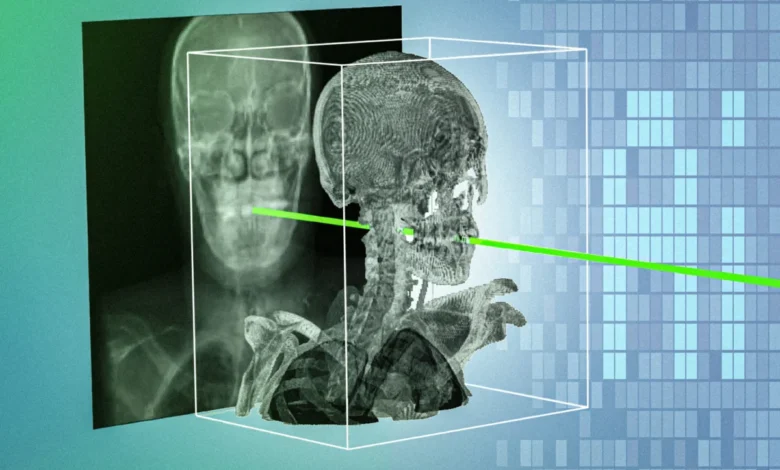
Researchers at the Massachusetts Institute of Technology (MIT), in collaboration with a network of clinical institutions, have developed a groundbreaking computational technique designed to rapidly and accurately match real-time X-rays captured during surgery with a patient’s preoperative three-dimensional medical scans. The novel system, designated as xvr (X-ray volume registration), overcomes long-standing technological hurdles in medical imaging by achieving sub-millimeter precision in a matter of seconds. By bridging the gap between flat, two-dimensional fluoroscopic imagery and volumetric data such as computed tomography (CT) and magnetic resonance imaging (MRI), the technology is poised to fundamentally transform the safety, precision, and accessibility of minimally invasive medical procedures.
The findings, published in the prestigious journal Nature, arrive at a critical juncture for modern medicine. Minimally invasive procedures—ranging from endovascular catheter insertions for stroke treatment to complex pediatric orthopedic surgeries—rely heavily on fluoroscopy, a type of continuous X-ray imaging, to guide clinicians through microscopic incisions. While these techniques spare patients the trauma of open surgery, they present a profound visual challenge: operators must navigate complex anatomical pathways using flat, two-dimensional projections that lack depth perception.
Bridging the 2D-3D Divide in the Operating Room
For decades, navigating the human body through real-time X-rays has required exceptional spatial reasoning and years of specialized clinical training. Because standard X-rays compress three-dimensional anatomical structures into a two-dimensional plane, determining the exact location, orientation, and trajectory of a surgical tool—such as a catheter, guidewire, or endoscope—remains notoriously difficult. Miscalculations can lead to vessel perforations, prolonged operating times, and severe complications.
To mitigate these risks, clinicians frequently attempt to manually register or align real-time X-rays with the patient’s preoperative 3D imaging data. This preparatory mapping allows physicians to visualize surgical instruments in the context of surrounding organs, bones, and blood vessels. However, manual registration is a laborious, highly subjective process. It typically requires a clinician to manually adjust digital overlays by entering numerical coordinates or clicking on anatomical landmarks on a computer screen while the patient is on the operating table. In high-stakes environments, such as acute stroke care or trauma surgery, the minutes spent performing manual alignments are an unacceptable luxury.
Over the past decade, artificial intelligence and machine learning researchers have attempted to automate this alignment process. Yet, these early AI systems faced a fundamental limitation: human anatomical diversity. A deep-learning model trained to align images for one patient often failed when confronted with the unique skeletal structures, pathological deformities, or varying body mass indices of another. Because large, high-quality annotated datasets of paired 2D X-rays and 3D scans are difficult to curate due to patient privacy laws and technical constraints, conventional neural networks struggled to generalize across diverse clinical populations.
The Genesis and Mechanics of the xvr Framework
To solve the generalization problem, the MIT-led research team abandoned the conventional paradigm of building a single universal AI model intended to work identically for every human being. Instead, they inverted the engineering approach, designing a system that tailors an individualized AI model specifically adapted to the anatomical architecture of each patient.
The newly developed xvr framework operates through a sophisticated, multi-step pipeline. First, the system ingests the patient’s preoperative 3D medical scan—whether a CT or an MRI. Utilizing a physics-based simulation of the X-ray imaging process, xvr synthesizes thousands of digitally reconstructed radiographs from a myriad of virtual angles. Generating approximately 1,000 synthetic images per second, the simulation relies entirely on the patient’s actual anatomical data rather than generative AI hallucinations, ensuring complete clinical fidelity and eliminating the risk of fabricated anatomical structures.
This patient-specific synthetic dataset is then used to train a dedicated neural network designed to align the patient’s real-time intraoperative X-rays with their volumetric scans. While this hyper-localized training approach yields unprecedented sub-millimeter accuracy, training a deep neural network completely from scratch for every incoming patient would theoretically require roughly 12 hours of computation—rendering it useless in an emergency operating room.
To overcome this latency barrier, the researchers engineered a breakthrough foundation model. Utilizing whole-body 3D medical scans from more than 2,000 diverse patients—spanning a wide demographic range, multiple imaging modalities, and various anatomical regions—the team pretrained a versatile foundational AI. When deployed for a new patient, this pretrained foundation model leverages the patient’s specific scan and rapidly adapts in approximately five minutes. Once adapted, it performs real-time 2D/3D image registration in mere seconds, matching the pristine accuracy of a model trained from scratch over half a day.
Rigorous Validation Across Diverse Clinical Environments
The research team subjected the xvr framework to rigorous validation using the largest known dataset of real-world 2D/3D registrations assembled to date. Incorporating multi-center data from five distinct hospitals, the evaluation tested the algorithm across dozens of different bone structures and organ systems in both adult and pediatric patient populations.
According to the study’s empirical metrics, xvr outperformed existing state-of-the-art artificial intelligence registration methods by an order of magnitude. Its superior robustness and velocity demonstrated that the system can maintain sub-millimeter precision across challenging anatomical geometries and variable imaging conditions, meeting the stringent timing requirements of emergency surgical interventions.
Lead author Vivek Gopalakrishnan, a postdoctoral researcher in the MIT Computer Science and Artificial Intelligence Laboratory (CSAIL) and a recent graduate of the Harvard-MIT Program in Health Sciences and Technology, emphasized the profound societal implications of democratizing access to precision imaging.
"A majority of Americans live more than an hour away from a center that can perform noninvasive procedures, like emergency stroke interventions," Gopalakrishnan noted. "An hour in stroke time is incredibly substantial. Making these procedures easier by combining 2D and 3D information enables these types of highly specialized life-saving procedures to be more accessible to much broader parts of the population."
A Collaborative Effort Rooted in Interdisciplinary Science
The development of xvr represents a deeply collaborative milestone bridging computer science, radiology, neurosurgery, and pediatrics. Gopalakrishnan was joined by co-senior authors Polina Golland, the Sunlin and Priscilla Chou Professor of Electrical Engineering and Computer Science at MIT, principal investigator in CSAIL, and leader of the Medical Vision Group; and Neel Dey, a former CSAIL postdoc who now serves as an investigator at Harvard Medical School and Massachusetts General Hospital.
The broader interdisciplinary team includes David-Dimitris Chlorogiannis, a researcher and clinician at Harvard Medical School; Andrew Abumoussa, a neurosurgeon at St. Luke’s Marion Bloch Neuroscience Institute; Anna M. Larson, a pediatric clinician at Shriners Children’s Hospital; Nazim Haouchine, an assistant professor of radiology at Harvard and Brigham and Women’s Hospital; Darren B. Orbach, a physician and scientist at Boston Children’s Hospital; and Sarah Frisken, an associate professor of radiology at Harvard.
Broader Implications for Robotic Surgery and Emergency Medicine
Beyond its immediate application in standard catheter- and needle-based interventions, the introduction of xvr holds substantial promise for the rapidly expanding field of surgical robotics. Modern robotic surgical platforms frequently rely on precise spatial mapping to execute pre-programmed trajectories or assist human operators with tremor filtration and boundary constraints. Integrating xvr into robotic surgical ecosystems could enhance autonomous navigation, allowing robotic arms to dynamically adjust to internal anatomical shifts during operations.
Furthermore, the technology addresses a persistent bottleneck in healthcare equity: the centralization of advanced procedural expertise. Highly complex minimally invasive interventions are traditionally restricted to major academic medical centers equipped with advanced visualization suites and specialized interventional radiologists. By translating grainy, two-dimensional fluoroscopy into intuitive, high-resolution augmented perspectives derived from preoperative scans, xvr lowers the cognitive barrier to entry for clinicians practicing in community hospitals and regional medical centers.
Future Outlook and Path to Clinical Translation
Following two years of meticulous algorithmic development and retrospective validation, the MIT team is shifting its focus toward translational development and commercial deployment. The researchers are actively collaborating with surgical robotics companies and major hospital clinical groups to adapt the xvr framework into deployable software and hardware navigation tools suitable for routine clinical workflows.
Near-term research objectives for the MIT group include optimizing the computational pipeline to achieve even faster real-time processing speeds, conducting prospective clinical trials to further verify reliability under unpredictable operating conditions, and expanding the underlying algorithms to seamlessly manage complex dynamic environments, such as soft tissues undergoing continuous deformation from cardiac and respiratory motion.
Funding for this foundational research was provided, in part, by the National Institutes of Health (NIH), the MIT CSAIL-Wistron Program, the MIT-IBM Computing Research Lab, the MIT Jameel Clinic, the MIT Health and Life Sciences Collaborative, and the Chou Family Transformative Research Fund. As these technological refinements progress from academic laboratories into commercial medical devices, xvr stands to redefine the visual standards of modern surgery, offering clinicians an unprecedented window into the human body when every second counts.
Related posts: