Revolutionary AI System Transforms Minimally Invasive Surgery by Rapidly Aligning 2D X-Rays with 3D Scans

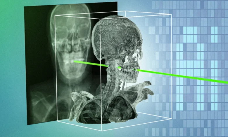
The landscape of modern medical interventions stands on the brink of a profound transformation following a breakthrough by researchers at the Massachusetts Institute of Technology (MIT) and a consortium of leading clinical institutions. The team has engineered an advanced artificial intelligence technique capable of bridging the gap between two-dimensional, real-time X-ray imagery and pre-procedural three-dimensional medical scans with unprecedented speed and sub-millimeter precision. Named xvr—short for X-ray volume registration—the system addresses a long-standing spatial orientation hurdle in minimally invasive medicine. By translating flat imaging into comprehensive navigational maps within minutes, this innovation promises to drastically reduce procedural complications, democratize access to emergency treatments, and supercharge the accuracy of surgical robotics.
The technological leap comes at a critical time for healthcare systems worldwide, where precision and speed dictate patient survival rates, particularly in time-sensitive emergencies like acute ischemic strokes or severe vascular blockages.
The Spatial Challenge in Minimally Invasive Interventions
For decades, the standard of care for many cardiovascular, neurological, and pediatric procedures has shifted away from traditional open surgeries toward minimally invasive techniques. Surgeons rely on tiny incisions to thread delicate medical devices—such as catheters, guidewires, and endoscopes—through complex vascular and anatomical pathways. To navigate these unseen internal corridors, clinicians depend on real-time fluoroscopy, a continuous X-ray imaging method that provides a live stream of the procedure.
However, standard X-rays are inherently two-dimensional flat projections. They capture three-dimensional anatomical structures by collapsing them onto a single plane, stripping away depth perception. Consequently, determining the exact three-dimensional orientation and trajectory of a surgical tool relative to critical organs, blood vessels, or tumors remains an extraordinary mental and technical challenge. Surgeons must interpret grainy, overlapping shadows, an interpretive skill that typically requires years of rigorous clinical training to master.
To mitigate the risk of misplacement, which can lead to catastrophic hemorrhages, vascular perforations, or damage to healthy tissue, clinicians frequently attempt to manually register or align real-time X-rays with preoperative diagnostic scans, such as volumetric Computed Tomography (CT) or Magnetic Resonance Imaging (MRI) datasets. Manual registration, however, is notoriously cumbersome. It requires clinicians to interrupt workflow, manually input coordinates, or click specific anatomical landmarks on a monitor to estimate tool positioning. In high-stakes emergency environments, where every second counts, manual alignment is simply too slow to be practical.
The Limitations of Prior Generative AI and Deep Learning Attempts
Recognizing the bottlenecks of manual registration, computer scientists and medical researchers have spent years attempting to harness artificial intelligence to automate the 2D/3D alignment process. Despite numerous efforts, previous deep-learning algorithms have suffered from a fundamental flaw: biological variability.
Human anatomy varies wildly across populations in terms of skeletal geometry, organ placement, and pathology. An AI model trained to recognize anatomical landmarks on one patient frequently fails when applied to another with a different body habitus, age, or underlying disease state. Furthermore, the medical imaging field suffers from a acute shortage of densely annotated, high-quality multi-modal training data, making it exceptionally difficult to train generalized deep-learning networks that can reliably handle the entire spectrum of human physiological diversity.
Rather than chasing the elusive goal of a universal AI model that attempts to process every human body identically, the MIT-led research team altered their foundational philosophy. They shifted the paradigm toward a patient-specific machine learning framework, optimizing the computational power to adapt exclusively to the individual lying on the operating table.
The Engineering Mechanics Behind xvr: Physics Meets Machine Learning
The xvr system bypasses the limitations of generalized models by adopting a two-pronged strategy: rigorous physics-based simulation paired with a rapid-adaptation foundation model.
The process begins immediately after a patient undergoes a standard preoperative 3D scan, such as a CT or MRI. The xvr framework takes this volumetric data and feeds it into a high-throughput, physics-based simulation engine. Within seconds, the system models the physical interaction of X-ray beams passing through tissues, generating up to 1,000 synthetic, digitally reconstructed radiographs per second from virtually every conceivable viewing angle.
Crucially, because these training images are generated via deterministic physics simulations derived directly from the patient’s own diagnostic scans rather than pure generative estimation, the risk of AI hallucination is entirely eliminated. There is no guesswork involved; the synthetic images strictly reflect the patient’s unique anatomical reality.
These thousands of patient-specific synthetic views are then utilized to train an alignment algorithm. While training an accurate model from scratch would normally take roughly 12 hours—making it useless for acute care—the researchers solved this time barrier by pretraining a massive foundation model. Utilizing a diverse dataset encompassing whole-body 3D scans from more than 2,000 patients across various age demographics, imaging modalities, and clinical centers, the team trained the core system to understand spatial transformations broadly.
When a new patient arrives, the pretrained foundation model leverages the patient-specific synthetic data generated by the physics simulator to adapt itself to the individual’s anatomy in approximately five minutes. Once calibrated, the system performs real-time 2D/3D X-ray registration in a matter of seconds with sub-millimeter precision.
Rigorous Validation Across Diverse Clinical Domains
To verify the robustness and reliability of xvr, the research team conducted the largest evaluation of its kind using real clinical data. The validation dataset incorporated multi-institutional records spanning five major hospitals, covering dozens of distinct skeletal structures and organ systems in both adult and pediatric cohorts.
The results, published in the prestigious journal Nature, demonstrated that xvr outperformed existing state-of-the-art AI registration methods by an order of magnitude. Its performance remained exceptionally stable across a wide spectrum of physiological variations, challenging image qualities, and procedural types.
The implications of this performance leap extend far beyond standard hospital operating rooms. According to study lead author Vivek Gopalakrishnan, a postdoctoral researcher in the MIT Computer Science and Artificial Intelligence Laboratory (CSAIL) and recent graduate of the Harvard-MIT Program in Health Sciences and Technology, the technology could fundamentally alter healthcare equity and emergency response logistics.
"A majority of Americans live more than an hour away from a center that can perform noninvasive procedures, like emergency stroke interventions," Gopalakrishnan noted. "An hour in stroke time is incredibly substantial. Making these procedures easier by combining 2D and 3D information enables these types of highly specialized life-saving procedures to be more accessible to much broader parts of the population."
Collaborative Chronology and Institutional Support
The development of xvr represents the culmination of years of multidisciplinary collaboration between computer scientists, bioengineers, and practicing clinicians.
The core research was spearheaded within MIT’s CSAIL Medical Vision Group, directed by Polina Golland, the Sunlin and Priscilla Chou Professor of Electrical Engineering and Computer Science and co-senior author of the study. The team worked alongside Neel Dey, a former CSAIL postdoc now serving as an investigator at Harvard Medical School and Massachusetts General Hospital, who shares co-senior authorship.
The clinical validation phases relied heavily on real-world insights from a broad coalition of medical professionals, including David-Dimitris Chlorogiannis of Harvard Medical School; Andrew Abumoussa, a neurosurgeon at St. Luke’s Marion Bloch Neuroscience Institute; pediatric clinician Anna M. Larson of Shriners Children’s Hospital; Nazim Haouchine, assistant professor of radiology at Harvard and Brigham and Women’s Hospital; pediatric neuroradiologist and physician-scientist Darren B. Orbach of Boston Children’s Hospital; and Sarah Frisken, associate professor of radiology at Harvard.
This extensive research effort received financial backing from several prominent public and private entities, including the National Institutes of Health (NIH), the MIT CSAIL-Wistron Program, the MIT-IBM Computing Research Lab, the MIT Jameel Clinic, the MIT Health and Life Sciences Collaborative, and the Chou Family Transformative Research Fund.
Future Outlook: Commercialization, Robotics, and Real-Time Deployment
With the foundational algorithm successfully validated in retrospective studies, the MIT research group is shifting its focus toward translational development and clinical integration.
The primary technical objective moving forward is to optimize the software pipeline for ultra-fast, real-time edge deployment during live surgeries. Additionally, the team is working to expand the capabilities of xvr to manage more complex, dynamic clinical scenarios, such as tracking highly deformable or continuously moving body parts—including the beating heart and expanding lungs—where anatomical deformation adds another layer of spatial complexity.
Furthermore, the researchers are actively engaging with the medical technology sector. By partnering with surgical robotics manufacturers and hospital clinical groups, the team aims to embed xvr directly into next-generation robotic surgical navigation consoles and mobile C-arm X-ray machines.
"For the past two years, we’ve been carefully developing this algorithm and validating it," Gopalakrishnan stated. "Now, we are collaborating closely with surgical robotics companies and clinical groups to turn this research into useful tools for navigation or deployment."
As regulatory pathways and clinical trials approach, xvr stands as a prime example of how patient-specific artificial intelligence can solve long-standing bottlenecks in clinical workflows, transforming raw data into life-saving precision.