Revolutionary AI Model E(3)-Pose Transforms Fetal MRI Scans to Detect Brain Abnormalities with Unprecedented Precision

Despite millions of years of human evolution, the intrauterine development of a fetus remains an extraordinarily complex biological sequence. Highly vulnerable to a multitude of genetic, environmental, and metabolic disruptions, the human gestational timeline can occasionally veer off course, giving rise to developmental abnormalities. Among the earliest anatomical structures to form—and one that continues its intricate structural maturation long after birth—is the human brain. Identifying pathologies within this vital organ before birth is critical for early clinical intervention, yet the traditional diagnostic pipeline is fraught with technical hurdles.
When a routine prenatal ultrasound yields ambiguous findings or flags potential structural anomalies, attending physicians typically order a fetal magnetic resonance imaging (MRI) scan to secure a definitive diagnosis. However, conventional imaging modalities are not without imperfection. Movement inside the womb, technical limitations, and the inherent difficulties of scanning a dynamic environment can lead to missed diagnoses, obscured pathologies, or inconclusive results, leaving expectant parents and medical teams navigating a high-stakes information vacuum.
To surmount these clinical limitations, a collaborative team of researchers hailing from the Massachusetts Institute of Technology (MIT), Boston Children’s Hospital, and the French National Institute for Research in Digital Science and Technology (Inria) has developed an innovative deep-learning model. Entitled E(3)-Pose, the system is engineered to track and image a fetus’s head dynamically while it is in motion. By compensating for fetal movement in real time, the technology captures exceptionally high-resolution volumetric data that radiologists can meticulously examine for early signs of neurological disorders. The foundational research behind this breakthrough was formally presented at the 2026 European Conference on Computer Vision, marking a significant milestone in computational medical imaging.
The Mechanics of Fetal Motion and MRI Limitations
To understand the magnitude of the breakthrough achieved by E(3)-Pose, one must first examine the inherent physics and operational challenges of fetal MRI. Unlike adult patients who can be instructed to lie completely still during a diagnostic scan, a fetus inside the womb is in a state of nearly constant motion. It shifts positions, turns, kicks, and floats within amniotic fluid, completely beyond the conscious control of the mother.
During a standard fetal MRI procedure, the scanner acquires a series of rapid two-dimensional (2D) images, often referred to by technicians and radiologists as "slices." When stacked together via computational processing, these individual 2D planes are designed to reconstitute a comprehensive, high-resolution three-dimensional (3D) volume of the fetal head. However, if the fetus shifts its position while the scanner is acquiring data, the alignment is instantly compromised.
Each subsequent 2D slice may end up being captured from a completely different angle, spatial orientation, or coordinate plane. This spatial inconsistency creates severe geometric discrepancies, resulting in frustrating coverage gaps and distorted slice orientations within the final reconstructed image. For a radiologist tasked with identifying subtle neurological malformations—such as cortical dysplasia, ventriculomegaly, or abnormal neuronal migration—these gaps can obscure critical anatomical landmarks. Consequently, a physician may entirely miss a developing pathology or struggle to formulate an accurate diagnosis.
As Ramya Muthukrishnan, an MIT doctoral student and the first author of the research, explains, the clinical margin for error is razor-thin. "One of the problems with having the baby move is that the slices are all over the place," Muthukrishnan notes. She conducts her research within the laboratory of Polina Golland, the Sunlin (1966) and Priscilla Chou Professor of Electrical Engineering and Computer Science at MIT, who serves as a principal investigator at the MIT Abdul Latif Jameel Clinic for Machine Learning in Health (Jameel Clinic) and the MIT Computer Science and Artificial Intelligence Laboratory (CSAIL).
"There are very fine details that you need a very good volumetric picture of the brain to see," Muthukrishnan elaborates. "If you have coverage gaps, you can’t see critical regions, and you need to be able to see well to detect abnormal development."
Hardcoding Physics into Deep Learning Architecture
What sets E(3)-Pose apart from conventional machine learning architectures used in medical imaging is its foundational reliance on the fundamental laws of physics. Rather than forcing a standard neural network to blindly learn the physical properties of motion and spatial orientation strictly from raw training data—a common approach that often leads to errors when encountering out-of-distribution clinical scenarios—the researchers baked core physical principles directly into the model’s structural architecture.
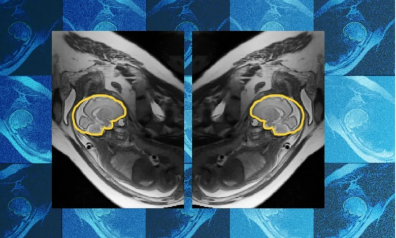
Specifically, the left-right anatomical symmetry inherent to the human fetal head is embedded directly into E(3)-Pose’s mathematical framework. This geometric equivariance ensures that the neural network does not need to waste computational power deducing basic biological constants; instead, it uses these invariant physical rules as a permanent compass. Consequently, the model can rapidly and accurately identify the exact orientation of the brain in real time, regardless of how erratically the fetus moves during the scanning process.
The path to developing this sophisticated model was far from straightforward. When Muthukrishnan embarked on her doctoral studies at MIT, her academic background was primarily rooted in computer science, with virtually no prior exposure to physics or the complex engineering principles governing MRI hardware. Transitioning from abstract algorithms to physical hardware deployment proved to be a baptism by fire.
"I was surprised when I had to go and implement this on the scanner," she recalls, reflecting on the grueling testing phase of her research. "I was not prepared for how many times I would crash the scanner, because putting things on hardware and getting it to work on hardware is a different beast than training a model to run on data on a GPU."
A Collaborative Clinical Journey
The genesis of E(3)-Pose was deeply influenced by real-world clinical observations. At the onset of her PhD program, Muthukrishnan spent time shadowing pediatric radiologists performing fetal MRI scans at Boston Children’s Hospital. During these clinical observations, she witnessed firsthand the profound frustration experienced by both patients and medical professionals.
Expectant mothers, already experiencing the anxieties associated with high-risk pregnancies, would endure physical discomfort lying motionless inside a confined MRI machine for upwards of an hour, only to have the scan aborted halfway through due to excessive fetal movement or maternal fatigue. Simultaneously, radiologists were left grappling with incomplete, low-quality datasets that failed to provide the necessary clarity to monitor fetal neurodevelopment effectively.
"Seeing frustration about it helped me decide what I wanted to do with my PhD, because it felt like this was a pain point," Muthukrishnan states.
Fortunately, she did not have to tackle these engineering challenges alone. A multidisciplinary team of clinicians, physicists, and computer scientists rallied around the project to bridge the gap between theoretical computer science and practical hospital implementation. Co-author Borjan Gagoski, an assistant professor of radiology at Harvard Medical School and a researcher at Boston Children’s Hospital, engineered a specialized MRI acquisition sequence. This custom sequence was vital for gathering the precise data required to train and validate E(3)-Pose, while Gagoski also worked closely with Muthukrishnan to troubleshoot the MRI hardware whenever software integration caused the system to crash.
Additionally, Aryn Lee, a research assistant at Boston Children’s Hospital, played a critical role in recruiting pregnant volunteers for the clinical study and assisting with the smooth acquisition of MRI scans under hospital protocols. The senior leadership team guiding the project included Elfar Adalsteinsson, an MIT professor of electrical engineering and computer science; Ellen Grant, the Käthe Beutler Professor of Pediatrics and professor of radiology at Harvard Medical School; and Polina Golland. Benjamin Billot, a researcher at Inria and a former postdoctoral fellow in Golland’s research group, provided essential early mentorship, helping Muthukrishnan integrate the foundational methodology he had previously developed.
Clinical Endorsements and Transformative Implications
The medical community has responded with enthusiasm to the successful validation of E(3)-Pose, praising its seamless integration of advanced mathematics and clinical utility.
"Ramya’s work elegantly combines geometric concepts like equivariance and symmetry with the power of deep learning to change how MRI scanners acquire the images, and to ultimately help the physicians detect and treat problems early in development," remarks Polina Golland. "It’s an impressive research project that is deep and fundamental in how it models image acquisition, with clear clinical impact."
Dr. Ellen Grant, who directs the Fetal Neonatal Neuroimaging and Developmental Science Center (FNNDSC) at Boston Children’s Hospital, emphasizes the administrative and patient-care advantages the technology introduces to modern obstetrical radiology. According to Grant, Muthukrishnan’s research "will automate fetal brain MRI, replacing today’s slow, manual process with faster and more comfortable scans for mothers, while also providing higher-quality images for radiologists to interpret."
By drastically reducing scan times and eliminating the need for repeat appointments caused by motion artifacts, E(3)-Pose promises to alleviate a significant source of psychological and physical stress for expectant mothers. Furthermore, by streamlining the workflow for radiologists, the technology could democratize access to high-fidelity fetal neuroimaging, ensuring that even subtle structural abnormalities are flagged well before birth.
Beyond the Womb: Future Horizons and Applications
While the immediate clinical application of E(3)-Pose is centered on third-trimester fetal brain MRI, the underlying technological framework possesses vast potential across multiple scientific and industrial domains. The mathematical principles governing E(3)-Pose’s ability to track non-rigid, moving objects from noisy, incomplete sensor data can readily be adapted to image other difficult-to-capture anatomical structures, such as beating hearts, rapidly moving joints, or vascular networks.
Moreover, the core architecture holds significant promise outside of medicine entirely. In fields like autonomous driving, where vehicle-mounted sensors must constantly interpret chaotic, real-world environments by processing noisy 3D data in real time to track moving pedestrians and vehicles, E(3)-Pose’s physics-informed approach could lead to more robust perception systems.
Despite these expansive possibilities, Muthukrishnan and her research collaborators remain focused on perfecting the technology for its most urgent medical application: expanding its capabilities downward into the second trimester of pregnancy. Currently, E(3)-Pose has been primarily trained and validated on data from third-trimester fetuses. Adapting the model for younger, smaller fetuses presents a formidable engineering challenge.
"Second-trimester fetuses are so small, they move around a lot more because there’s more space in the uterus," Muthukrishnan explains. "They’re more important to focus on because the earlier you detect brain abnormalities, the easier it is to intervene."
As research progresses toward second-trimester applications, the collaborative team is continuing to refine the model’s algorithms and expand its clinical trial datasets. The development of E(3)-Pose has been made possible through generous financial support from several key institutions, including the MIT Jameel Clinic, the MIT Takeda Program, the Chou Family Transformative Research Fund, the National Institute of Biomedical Imaging and Bioengineering, and the Eunice Kennedy Shriver National Institute of Child Health and Human Development.
Through the fusion of advanced artificial intelligence, rigorous physical modeling, and dedicated clinical collaboration, E(3)-Pose stands as a testament to the power of interdisciplinary research. By bringing unprecedented clarity to one of the most challenging frontiers in diagnostic medicine, this technology heralds a new era of prenatal care—one where early detection translates directly into a healthier start to life.